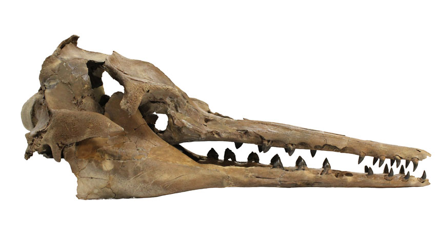
A roughly 27-million-year-old fossilized skull echoes growing evidence that ancient whales could navigate using high-frequency sound.
Discovered over a decade ago in a drainage ditch by an amateur fossil hunter on the South Carolina coast, the skull belongs to an early toothed whale. The fossil is so well-preserved that it includes rare inner ear bones similar to those found in modern whales and dolphins. Inspired by the Latin for “echo hunter,” scientists have now named the ancient whale Echovenator sandersi. “It suggests that the earliest toothed whales could hear high-frequency sounds,” which is essential for echolocation, says Morgan Churchill, an anatomist at the New York Institute of Technology in Old Westbury. Churchill and his colleagues describe the specimen online August 4 in Current Biology.
Modern whales are divided on the sound spectrum. Toothed whales, such as orcas and porpoises, use high-frequency clicking sounds to sense predators and prey.
Filter-feeding baleen whales, on the other hand, use low-frequency sound for long-distance communication. Around 35 million years ago, the two groups split, and E. sandersi emerged soon after.
CT scans show that E. sandersi had a few features indicative of ultrasonic hearing in modern whales and dolphins. Most importantly, it had a spiraling inner ear bone with wide curves and a long bony support structure, both of which allow a greater sensitivity to higher-frequency sound. A small nerve canal probably transmitted sound signals to the brain. “Scientists have long suspected that early toothed whales could produce the high-frequency sounds needed for echolocation based on features on their skulls,” says Travis Park of Monash University in Melbourne, Australia. Previous work points to early toothed whales sensing those high frequencies. Park and his colleagues reported in April in Biology Letters the discovery of a 26-million-year-old lone ear bone showing signs of high-frequency hearing. But it wasn’t connected to a skull and, thus, couldn’t be tied to a specific whale species.
Tracing inner ear features in CT scans of 24 ancient and modern whales, including E. sandersi, plus two hippos, whales’ closest living relatives, Churchill’s team ups the ante. Because primitive versions of the bony spiral and nerve canal appeared before the first known toothed whale, the researchers suggest that rudimentary high-frequency hearing might have emerged in the common ancestor of toothed and baleen whales at least 43 million years ago. If so, baleen whales lost their high-frequency hearing at some point. Determining whether that’s truly the case requires more analysis and a wider array of fossil data, says Park, who’s unconvinced.
But there is growing consensus that the first toothed whales could hear and produce sounds at high-frequency ranges. The skull of a 28-million-year-old toothed whale also suggests that such animals could make high-frequency calls (SN: 4/19/14, p. 6). “The next step is to look at when their brains got big enough to process echolocation signals,” says Nicholas Pyenson, a paleobiologist at the Smithsonian’s National Museum of Natural History who was not affiliated with the study. “This is great, but there’s more to be done.”
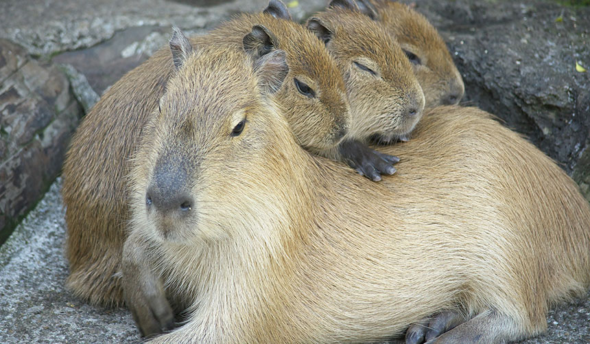
Capybaras, giant rodents native to South America, could become Florida’s next big invasive species, a biologist warned August 3 in Columbia, Mo., at the 53rd Annual Conference of the Animal Behavior Society.
“Capybaras have been introduced to northern Florida,” said Elizabeth Congdon of Bethune-Cookman University in Daytona Beach, Fla. And there are enough similarities to nutria — large invasive rodents that have caused havoc in many states — to warrant a closer look at the South American newcomers.
There are currently about 50 capybara loose in northern Florida. Now, that may not seem like an invasion, and it’s not — yet. But these animals are the world’s largest rodent, growing to 50 kilograms or more. In the wild, the semiaquatic animals live in social groups in forests where they can be near bodies of water, such as rivers, lakes or swamps. They are herbivores that can subsist on a wide variety of vegetation, from grass to tree bark. And they reproduce at a fair pace, producing an average of four, and up to eight, pups per litter.
Most people wouldn’t look at those characteristics and think “I want to own one of those animals,” but some have. Capybaras are one of the many exotic creatures that people have tried to turn into pets. (Owning one is legal in some states.) But the animals can get loose, or people may purposely release them when they no longer want to own a giant rodent.
A capybara (or 50) loose in the countryside or city is not automatically an invasive species. The difference between an invasive and a nonnative exotic is whether an organism is causing environmental or economic harm, or harm to human health.
Congdon and her undergraduate students have been studying the potential for capybaras to make that transition from exotic to invasive, and they have been looking for similarities to nutria. Those large rodents were first imported to the United States in the early 1900s; the animals were farmed for their fur in Louisiana. But they escaped — some were also purposely released as weed mitigators — and quickly established themselves in Louisiana’s many swamps. Efforts to control the animals, such as hunting, have largely failed.
Nutria, which are smaller than capybaras, reproduce at about the same rate as the giant rodents. But one of the things that have made nutria such a menace — their propensity to dig into riverbanks, levees and other places that can cause problems when the ground disintegrates — appears to be a trait they don’t share with capybaras. While coyotes and dogs are know to hunt nutria, it appears that nothing in the United States, other than a human, is big enough to kill a capybara. No animals here are equivalent to the capybara’s natural South American predators, which include anacondas, puma and jaguar, Congdon noted.
The state of Florida says only that a breeding population of capybaras “may exist,” but Congdon is pretty sure that there is one. In 1995, five animals escaped from a wildlife facility near Gainesville, and they are probably at least part of the source of those 50 capybaras now living in Florida. “Several sightings suggest they have been breeding,” Congdon said, including the finding of a juvenile capybara. Given the similarities to the nutria, and the ability of capybaras to adapt to a variety of habitats, including cities, “they might be able to make a go of it in the United States,” Congdon concluded.
But Congdon isn’t advocating that wildlife managers kill all the capybaras in Florida. The animals represent “an opportunity to study the process of invasion,” she said. Plus, a population in Florida would be a lot easier for her to access than the one she studied in Venezuela as a gradate student. “We want to keep them from spreading,” she said, “but can we please not kill them all so I can study them?”
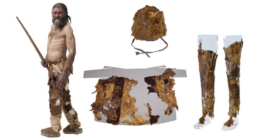
The 5,300-year-old Tyrolean Iceman, whose body was found poking out of a glacier in the Italian Alps in 1991, incorporated hides from at least five domesticated and wild animal species into his apparel, a new genetic study finds. Comparing mitochondrial DNA extracted from nine ancient leather fragments with DNA of living animals revealed the makeup of Ötzi’s clothes and a key accessory, says a team led by paleogeneticist Niall O’Sullivan. Mitochondrial DNA typically gets passed from mothers to their offspring. Little is known about what people wore during Ötzi’s time. The findings provide a glimpse into how ancient European populations exploited domesticated animals to make clothes and other items.
Ötzi’s coat consisted of hides from at least three goats and one sheep, the scientists report August 18 in Scientific Reports. This garment may have been periodically patched with leather from whatever animals were available, the team suggests.
Goats also provided skin for the Iceman’s leggings, the new analysis indicates.
A sheepskin loincloth and a shoelace derived from European cattle round out Ötzi’s attire made from domesticated animals.
As for wild animals, Ötzi wore a brown-bear cap and toted a quiver made from roe deer. It’s impossible to know if the ancient man attached any special meaning to brown bears, “but he may have been an opportunistic hunter or a scavenger,” says O’Sullivan, of University College Dublin and EURAC Research in Bolzano, Italy. A 2012 analysis of proteins from fur samples taken from Ötzi’s clothing identified sheep and a goatlike animal called a chamois as sources for the Iceman’s coat. A team led by biochemist Klaus Hollemeyer of Saarland University in Saarbrücken, Germany, also pegged goats and dogs or wolves as sources of skin for Ötzi’s leggings.
Disparities between Hollemeyer’s and O’Sullivan’s studies may stem from the two groups having sampled different parts of patchwork garments. In addition, the new report used advanced techniques for extracting and analyzing ancient DNA. That enabled O’Sullivan’s team to retrieve six complete mitochondrial genomes from Ötzi’s leather belongings.
O’Sullivan’s investigation “opens a new field of potential identification procedures for mammalian species in ancient leathers and furs,” Hollemeyer says.
A roughly 4,200-year-old legging found in the Swiss Alps in 2004 also features goat hide. Mitochondrial DNA extracted from that garment came from an ancient line of European goats that has largely been replaced by a genetically distinct goat population, a team led by archaeologist Angela Schlumbaum of the University of Basel in Switzerland reported in 2010.
The Swiss legging was found with pieces of bows and arrows, woolen clothes and many other artifacts where an ice patch in a mountain pass had partly melted. No human bodies have been found there.
“Possibly, goat leather was most comfortable” as legging material, says University of Bern archaeologist Albert Hafner, a coauthor of the Swiss legging study. “Modern leather trousers often use goat as well.”
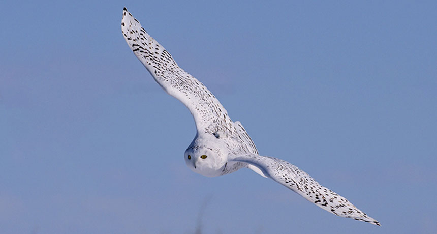
White, fierce and fluffy, snowy owls are icons of Arctic life. But some of these owls are not cool with polar winters.
Every year, part of the population flies south to North American prairies. Ornithologists thought those birds fled the Arctic in desperation, haggard and hungry. But the prairie owls are doing just fine, researchers report August 31 in The Auk: Ornithological Advances.
Over 18 winters, wild snowy owls caught and banded in Saskatchewan, Canada — one of the species’ southerly destinations — were 73 percent heavier than emaciated owls in rescue shelters. Females were heavier and had more fat than males, and adults were in better condition than youngsters. But regardless of age or sex, most snowy owls that made the journey south were in relatively good health.
That means southern winters may not be such a desperate move after all. Prairies are probably just a normal wintering ground for some of the Arctic snowy owl population, the researchers say. Snowbirds, indeed.
Philae has been found, nestled in a shadowy crevice on comet 67P/Churyumov-Gerasimenko. The comet lander, lost since its tumultuous touchdown on the comet on November 12, 2014, turned up in images taken by the Rosetta orbiter on September 2.
Philae is on its side with one leg sticking out into sunlight. Its cockeyed posture probably made it difficult for Philae to reliably get in touch with Rosetta, explaining why scientists had trouble reestablishing communication. The discovery came about a month before the end of the Rosetta mission; the orbiter was scheduled to land on the comet on September 30and then shut down.
Philae spent just a few days transmitting data from the comet’s surface (SN: 8/22/15, p. 13). It had a rough landing, bouncing twice before stopping. Sitting in the shadow of a cliff, Philae was unable to use solar power to recharge its battery. Rosetta picked up intermittent communication in June and July 2015. Since January, temperatures on the comet have been too chilly for Philae’s electronics; scientists stopped listening for radio signals in July.
Sea ice around the North Pole has reached its second-lowest low on record, tying with 2007, scientists at NASA and the National Snow and Ice Data Center announced September 15.
Arctic sea ice reached its expected low point for the year on September 10, bottoming out at an area of 4.14 million square kilometers. That’s well below the 1981 through 2010 average of 6.22 million square kilometers, though above the record-lowest extent of 3.39 million square kilometers, set in 2012.
The silver-medal finish came after a summer of relatively cool temperatures, cloudy skies and stormy weather — conditions that typically limit sea ice shrinkage. The lack of ice probably arose from a poor starting position: The melt season began with the smallest maximum sea ice extent on record.
Shrinking sea ice can speed up warming, threaten Arctic species and spread pollution.
Qian Chen, 30 Materials scientist University of Illinois
The SN 10 In a darkened room, bathed in the glow of green light, materials scientist Qian Chen watches gold nanorods dance. They wiggle across a computer screen displaying real-time video from a gigantic microscope — a tall, beige tube about as wide as a telephone pole.
Chen has observed these and other minuscule specks of matter swimming, bumping into one another and sometimes organizing into orderly structures, just like molecules in cells do. By pioneering the design of new biologically inspired materials, she’s exploring what it means to be “alive.” Next, Chen wants to get an up-close and personal view of cellular molecules themselves: the nimble, multitasking proteins that work day and night to keep living organisms running.
At age 30, Chen is already racking up high-profile publications and turning some far-out ideas into reality. Her ultimate goal: To mimic the machinery that living cells have already perfected. To create life, or something like it, out of nonliving materials.
“If you can see it, you can start to understand it,” Chen said when I visited her lab at the University of Illinois at Urbana-Champaign earlier this year. “And if you understand it, you can start to control it.”
Chen didn’t always want to be a scientist. Growing up in China, she imagined one day becoming a writer. In middle school, she wrote an award-winning story about a girl who figures out how to repair the ozone layer. “My idea was to get some material that can be stretched, like the skin of the balloon,” Chen says. Her interest in inventing new and unusual materials took off years later, in the United States. After graduating from college in China in 2007 — Chen was the first in her family to do so — she headed to Illinois to work with materials scientist Steve Granick.
From the start, Chen stood out. “She made hard things look easy,” says Granick, now at the Ulsan National Institute of Science and Technology in South Korea. He recalls one experiment in particular, when Chen performed a feat some scientists thought impossible: She got thousands of tiny beads to form an open and orderly two-dimensional structure — all by themselves.
Chen had been studying colloidal particles, microscopic specks roughly a micrometer in size. People normally think of these particles as a component of paint, not all that interesting.
But Chen had the idea to cover the particles with a kind of sticky coating that acted something like Velcro. When the particles bumped into one another, they stuck together. At first, “It looked like a mess, like a failed experiment,” says Granick. “Most graduate students would have just chalked it up to a mistake and gone home.”
After a day of knocking around in solution, sticking together and tearing apart, the particles finally settled into something stable. The special coating and the way Chen applied it (capping the top and bottom of each particle) led to a “kagome lattice,” something sort of like a honeycomb. Never before had scientists coaxed colloidal particles into such an open, porous framework. Usually, the particles pack together more tightly, like apples stacked on the shelf at a grocery store, Chen says. That work led in 2011 to a publication in Nature: “Directed self-assembly of a colloidal kagome lattice.” A week earlier, Chen and Granick had published a different paper in Science, “Supracolloidal reaction kinetics of Janus spheres,” about particles that self-assemble into a twisting chain, or helix. At the time, Chen was 24.
“Her work is at the leading edge,” says Penn State chemist Christine Keating. “She’s so full of enthusiasm for science, and energy and creative ideas.”
Exactly how such particles might one day be used is still anybody’s guess. Some researchers envision self-assembling materials building smart water filters or adaptable solar panels that change shape in response to the sun. But the full range of possibilities is hard to fathom. Chen is “trying to invent the rules of the game,” Granick says. “She’s laying the groundwork for future technologies.”
Her next big focus will take her field from self-assembly 101 to the master class level, by mimicking how biological molecules behave. But first she has to see them in action.
Into the cell In 2012, Chen traveled west to the University of California, Berkeley to work with National Medal of Science winner Paul Alivisatos on a new microscopy technique.
Scientists today can view the details of proteins and DNA close up under a microscope, but the results are typically still-life images, frozen in time. It’s harder to get action shots of proteins morphing in their natural, fluid world. That view could unveil what roles different protein parts play.
Even a technique that won its developers a Nobel Prize in 2014 (SN: 11/2/14, p. 15) — it relies on fluorescent molecules to illuminate a cell’s moving parts — can’t always reveal the intricacies of proteins, Chen says. They’re just glowing dots under the microscope. Imagine, for example, looking at a dump truck from an airplane window. You can’t see how the truck actually works, how the pistons help lift the bed and the hinges open the tailgate.
“I use this as inspiration,” Chen says, grabbing her laptop and starting up a video that may well be the fantasy of anyone exploring biology’s secret world. The computer animation shows molecules whizzing and whirling deep inside a cell. Gray-green blobs snap together in long chains and proteins haul giant, gelatinous bags along skinny tracks. No one yet has gotten a view as clear as this hypothetical one, but a technique Chen is now helping to develop at Illinois could change that.
It’s called liquid-phase transmission electron microscopy, and it’s a slick twist on an old method. In standard TEM, researchers create subnanometer-scale images by shooting an electron beam through samples placed in a vacuum. But samples have to be solid — still as stone — because liquids would evaporate.
By sandwiching beads of liquid between thin sheets of graphene, though, Chen gets around the problem. It’s like putting droplets of water in a plastic baggie. The liquid doesn’t dry up, so researchers can observe the particles inside jittering around. Chen has used the technique to see gold nanorods assembling tip-to-tip and DNA-linked nanocrystals moving and rotating in 3-D. Now, she may be on the verge of a big advance.
With liquid-phase microscopy, Chen is attempting to see cellular machinery with a clarity no scientist has achieved before. She is cautious about revealing too many details. But if Chen succeeds, she may be on her way to cracking the code that links biological structure to function — figuring out the parts of a protein, the pistons and hinges, that let it do its specific job. Knowing the structural building blocks of life, she says, will help scientists create them — and everything they can do — out of artificial materials.
“We’re not there yet,” Chen says, “but that’s the big dream.”
A baby boy born on April 6 is the first person to be born from a technique used to cure mitochondrial diseases, New Scientist reports.
The child’s mother carries Leigh syndrome, a fatal disease caused by faulty mitochondria. Mitochondria generate most of a cell’s energy and perform other functions that keep cells healthy. Each mitochondria has a circle of DNA containing 37 genes needed for mitochondrial function. A mutation in one of those genes causes Leigh syndrome. The woman herself is healthy, but previously had two children who both died of Leigh syndrome.
John Zhang, a fertility doctor at New Hope Fertility Center in New York City, and colleagues transferred a structure called the spindle with chromosomes attached to it from one of the woman’s eggs into a healthy, empty donor egg. The resulting egg was then fertilized with sperm from the woman’s husband. The procedure was done in Mexico.
The technique, called spindle nuclear transfer, is one of two ways of creating “three-parent babies” to prevent mitochondrial diseases from being passed on. Such three-parent babies inherit most of their DNA from the mother and father, but a small amount from the donor. Other three-parent children who carried mitochondria from their mothers and from a donor were born in the 1990s, but the baby boy is the first to be born using a nuclear transfer technique. Zhang and colleagues will report the successful birth October 19 in Salt Lake City at the American So
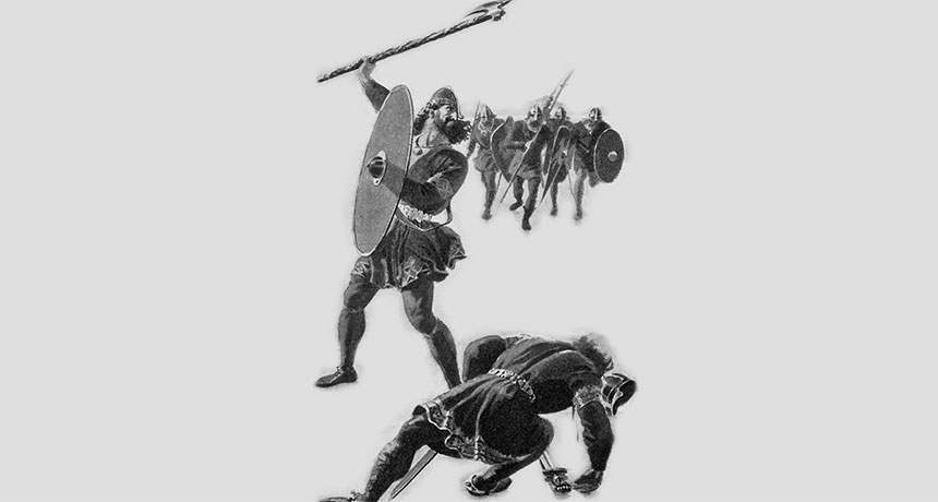
Murder was a calculated family affair among Iceland’s early Viking settlers. And the bigger the family, the more bloodthirsty.
Data from three family histories spanning six generations support the idea that disparities in family size have long influenced who killed whom in small-scale societies. These epic written stories, or sagas, record everything from births and marriages to deals and feuds.
Iceland’s Viking killers had on average nearly three times as many biological relatives and in-laws as their victims did, says a team led by evolutionary psychologist Robin Dunbar of the University of Oxford. Prolific killers responsible for five or more murders had the greatest advantage in kin numbers, the scientists report online September 20 in Evolution and Human Behavior. Particularly successful killers chose their victims carefully, knowing that their large families would deter revenge attacks by smaller families of the slain, the researchers contend. Those killings were motivated by land grabs, they suspect. One-time killers tended to have only slightly bigger families than those of their victims; insults or goading possibly prompted those murders. Strikingly, around 18 percent of all men mentioned in the sagas were murdered. Similarly high homicide rates, mainly due to cycles of revenge killing between feuding families, have been reported for some modern hunter-gatherer and village-based societies ( SN Online: 9/27/12 ). Lethal raids by competing groups may go back 10,000 years or more ( SN: 2/20/16, p. 9 Murder rates rise in the absence of central authorities that enforce social order, Dunbar proposes. “The real issue is not that there were so many murders among Icelandic Vikings, but that murders were carefully calculated based on knowing whether one had a sufficient family advantage to take the risk.”
That idea relates to mathematical formulas of fighting strength developed during World War I by British engineer Frederick Lanchester. One of Lanchester’s laws calculates that the fighting advantage of a larger group over a smaller group grows disproportionately as the disparity in the size of war parties increases. That rule also holds for family-size differences in small-scale societies, such as Icelandic Vikings, Dunbar’s group concludes.
Tests of the possibility that greater kin numbers encourage lethal attacks in preindustrial groups, such as the Vikings, are rare, says Oxford evolutionary biologist and political scientist Dominic Johnson, who did not participate in the new study. Johnson has reviewed evidence suggesting that humans, chimps and social hunters such as wolves have evolved ways to monitor group sizes and launch attacks when they can gang up on a few opponents.
Dunbar and his colleagues studied three Icelandic family sagas covering events from around 900 to 1100. Iceland’s first settlers arrived from Scandinavia and northern Europe in the late 800s (SN: 5/14/16, p. 13).
The sagas contained information about events, including feuds and murders, involving 1,020 individuals. For everyone mentioned, the researchers identified a network of biological and in-law relationships.
Under Norse law, a murder entitled a victim’s relatives to compensation, either via a revenge murder or blood money. Icelandic sagas describe the importance of avenging murdered relatives to save face and prevent further attacks, regardless of family size.
In the three sagas, a total of 66 individuals caused 153 deaths; two or more attackers sometimes participated in the same killing. No killers were biologically related to their victims (such as cousins or closer), but one victim was a sister-in-law of her killer.
About two-thirds or more of killers had more biological kin on both sides of their families, and more in-laws, than their victims did.
Six men accounted for about 45 percent of all murders, each killing between five and 19 people. Another 23 individuals killed two to four people. The rest killed once. Frequent killers had many more social relationships, through biological descent and marriage, than their victims did, suggesting that they targeted members of families in vulnerable situations, the researchers say.
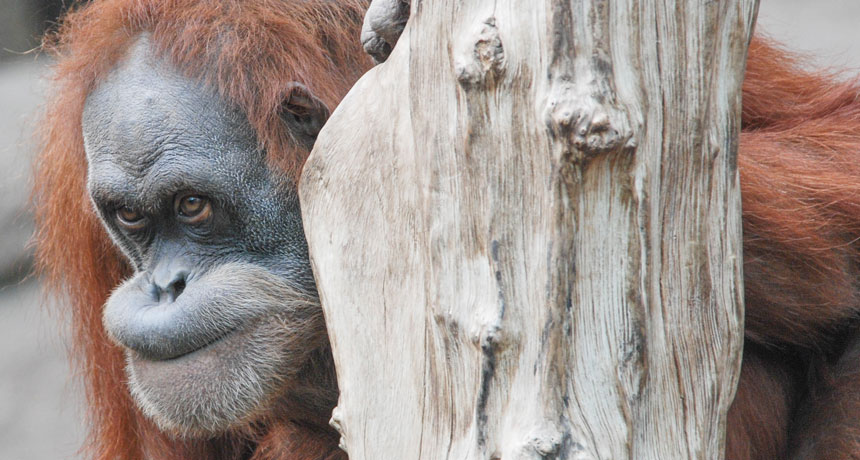
Apes understand what others believe to be true. What’s more, they realize that those beliefs can be wrong, researchers say. To make this discovery, researchers devised experiments involving a concealed, gorilla-suited person or a squirreled-away rock that had been moved from their original hiding places — something the apes knew, but a person looking for King Kong or the stone didn’t.
“Apes anticipated that an individual would search for an object where he last saw it, even though the apes knew that the object was no longer there,” says evolutionary anthropologist Christopher Krupenye. If this first-of-its-kind finding holds up, it means that chimpanzees, bonobos and orangutans can understand that others’ actions sometimes reflect mistaken assumptions about reality. Apes’ grasp of others’ false beliefs roughly equals that of human 2-year-olds tested in much the same way, say Krupenye of the Max Planck Institute for Evolutionary Anthropology in Leipzig, Germany, and his colleagues.
Considering their targeted gazes during brief experiments, apes must rapidly assess others’ beliefs about the world in wild and captive communities, the researchers propose in the October 7 Science. Understanding the concept of false beliefs helps wild and captive chimps deceive their comrades, such as hiding food from those who don’t share, Krupenye suggests.
Experiments included 41 apes — 19 chimps, 15 bonobos and seven orangutans. These animals had been born in captivity and lived in open enclosures at research centers in Germany and Japan. Apes watched two short videos designed to grab their attention. In one, a person in a King Kong gorilla suit hides in one of two haystacks while a man watches. After the man leaves through a door, King Kong runs away. Then the man returns and looks for King Kong. In a second video, a man returns for a stone that King Kong stole from him and hid in one of two boxes while the man watched. During the man’s absence, however, King Kong runs off with the stone or, in another version, moves the stone from one box to the other.
A camera equipped with an eye-tracking sensor revealed that, when the man in these videos returned, apes usually looked first at where King Kong or the stone had initially been hidden. They also spent more time looking at those initial locations than at any other spots in the videos. Those behaviors indicate that the apes assumed the man would return to those same spots based on where he had last seen what he was looking for. Of 29 animals that viewed both videos, gazes of 23 indicated that they expected the man in one or both scenarios to hold a false belief, the researchers say. Krupenye’s team shows for the first time that a nonhuman animal can track others’ false beliefs, agrees psychologist Amanda Seed of the University of St. Andrews in Fife, Scotland. But it has yet to be demonstrated that apes, like humans, can act on such knowledge, say by hiding food from others, she adds. It’s also unclear whether, aside from knowing where an observer will look for an item, apes truly know that the object is no longer there, Seed says. Further experiments could see if apes express surprise upon seeing an observer find an item hidden in its original location after it had been moved, she suggests.
An ability to infer what others are thinking, dubbed “theory of mind” by psychologists (SN Online: 3/27/13), likely evolved in ancient ancestors of humans and apes, writes primatologist Frans de Waal of Emory University in Atlanta in the same issue of Science. Those ancestors lived in increasingly complex communities where it paid to predict accurately how others would behave, he proposes.
Yale University psychologist Laurie Santos isn’t so sure apes track false beliefs. Previous research has consistently indicated that no nonhuman animals monitor others’ beliefs, even on tasks similar to those used by Krupenye’s team, Santos says. In the new study, she adds, apes may have realized that an observer was ignorant about an object’s new location but not that he had false expectations about where to find it.
Krupenye disagrees. “The apes specifically anticipated that the actor in the video would search for an object where we humans know the actor falsely believed the object to be,” he says.